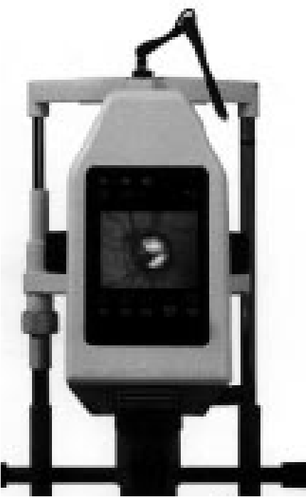
LVDT Use in a Scanning Laser Tomography Instrument
At one time, everyone feared a glaucoma test when visiting the optometrist – squirming in your chair while waiting for that sudden puff of air. That is until Laser Diagnostic Technologies, San Diego, Calif. designed a scanning laser system that uses a near infrared diode laser instead of an airstream for imaging and measurement of retinal topography. The light probe is nearly undetectable by the patient and images are acquired through undilated pupils.
The TopSS XL-2020 is a multiple purpose tomograph which offers several scan ranges and selectable placement of the image series. To maximize depth resolution for the retinal feature being measured, a lens is focused on an area of concentration. The position of the lens is precisely monitored by a Trans-Tek Model 0244-00000 DC-DC LVDT with a working range of ±1.00 inch. The XL-2020 includes three field of view choices allowing optimal lateral resolution for the target area.
The lens is positioned by a stepper motor, a main component of the imaging mechanism. As the stepper motor moves the lens into position, the Model 0244 LVDT sends a signal to the programmed software which processes this information to determine distance to the retinal area. The image produced is a result of the accurate LVDT measurement and relative positioning of the topographic feature. The transducer output is part of a continuous feedback loop in the scanner’s on-board logic control system.
A critical function of this eye examination is to compare results from previous tests. Such comparisons are used to detect any subtle changes and to help determine the appropriate medical treatment. The XL-2020 boasts reproducibility of 30 um in a pixel size of 11 um for each composite image. This is real data, not interpolated, giving objective information to support diagnostic decisions. With so much emphasis place on reproducibility, it is no wonder an LVDT was chosen as the position sensor. The repeatability of an LVDT often far surpasses its specification for linearity.
The final data is presented to the optometrist for analysis on a familiar Microsoft Windows platform. The XL-2020 provides this dependable data for the diagnosis and management of glaucoma with optic nerve head imaging and measurements. Other topographic features inspected by this system include macula holes, tumors, edema and more. The software offers eight different color scales, ten image enhancement routines and adjustable 3-D wire frame to assist with qualitative evaluation of the images. The XL-2020 automatically calculates reproducibility of topography measurements. Other capabilities include enhanced image visualization, volumetric analysis, and baseline and comparison images.
The Series 240 DC-DC LVDT provides accurate displacement measurement for strokes ranging from ±0.050” to ±3.00”. Convenience is a major selling point for this product line and is highlighted by complete signal conditioning electronics contained in a single integrated package for DC-in/DC-out operation.
Besides convenience, it was the stable and repeatable output signal that attracted design engineers at Laser Diagnostic to this DCDT. And for the medical industry, accuracy and resolution are key parameters that can never be compromised.